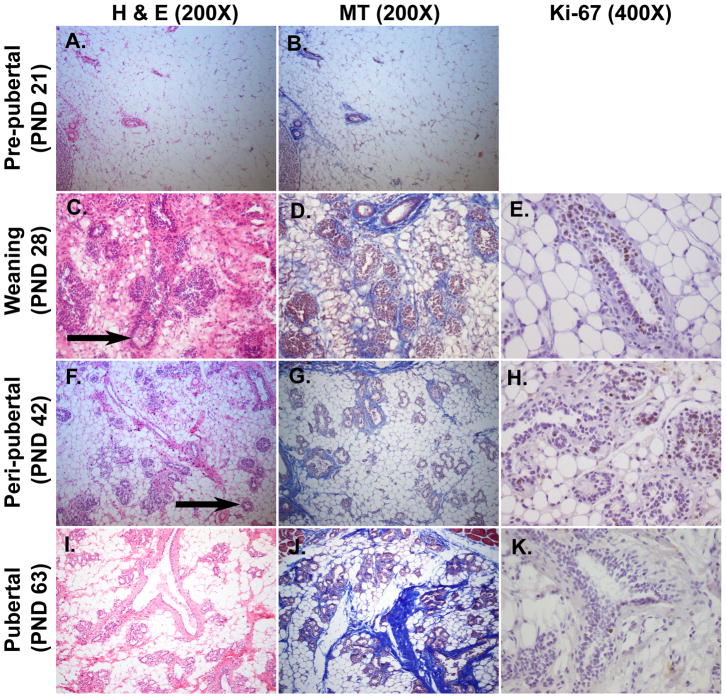
Figure 2. Histological features of mammary glands during early stage of development (PND 21-PND 63).
Panels A-C-F-I were stained with Hematoxylin and Eosin (H&E, 200X) and panels B-D-G-J with Mallory’s Trichrome (MT, 200X). Panels E-H-K were immunostained with Ki-67 antibody (400X). The second axillary mammary glands were dissected from female Sprague Dawley rats. A–B: The stromal compartment is predominant and filled with adipocytes. C-D: Ductal growth is visible as evidenced by formation of TEBs (arrow). E: TEBs are the sites of ductal elongation and branching and represent the sites of highest proliferation in the gland, as revealed by the positivity to immunohistochemistry for the marker of proliferation Ki-67. F–G: TEBs are characterized by a single layer of cap cells and multilayered pre-luminal body cells (arrow). H: Duct elongation is characterized as a proliferative process as revealed by immunohistochemistry for Ki-67 antibody. I-J: Glands are arranged into lobules of compound branched alveolar glands, separated by dense interlobular connective tissue and fat. K: Ki-67 is poorly expressed at pubertal stage, indicating that the exponential development is already occurred.

