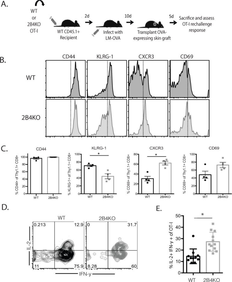
Figure 7. 2B4-mediated signals impact memory recall potential.

A, 104 WT or 2B4KO OT-I T cells were transferred into naïve B6 hosts, which were infected with 104 CFU LM-OVA 2d later. Animals received OVA-expressing skin grafts at day 10 post-transplant and were sacrificed five days later. B-C, Representative flow cytograms and summary data depicting the expression of CD44, KLRG-1, CXCR3, and CD69 on Thy1.1+ CD8+ OT-I T cells isolated from the spleen. D-E, Splenocytes were restimulated ex vivo with PMA and ionomycin and analyzed by ICS. Data shown in A–E are representative of 6-11 mice/group from 2 independent experiments. p = 0.0005.
