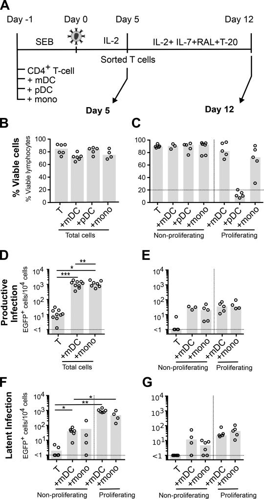
Figure 2. Latency in non-proliferating and proliferating CD4+ T-cells is maintained in a subset of cells in vitro.
Experimental conditions were as in Figure 1. A. A subset of sorted uninfected, non-proliferating and proliferating T-cells were cultured without stimulus for a further 7 days in the presence of IL-2, IL-7, HIV-1 fusion (T-20) and integrase inhibitors (L8 or Raltegravir; RAL) to measure stability of latent infection. Total cell culture viability was quantified by flow cytometry at B. day 5 (n=4–6); and C. day 12 (n=3–5). EGFP was quantified at D. day 5 (n=7–9); and E. day 12 (n=3–5) post-infection as a measure of productive infection. Latent infection was measured by quantification of EGFP expression following stimulation with anti-CD3/CD28 and IL-7 and in the presence of an integrase inhibitor at both F. day 5 (n=4–5); and G. day 12 (n=4–5). Columns represent the median; symbols represent results from individual donors. Significance was measured by a paired students T-test where n<5, and Wilcoxon signed-rank test where n≥5, *p≤0.05, **p≤0.005, ***p≤0.0005.

