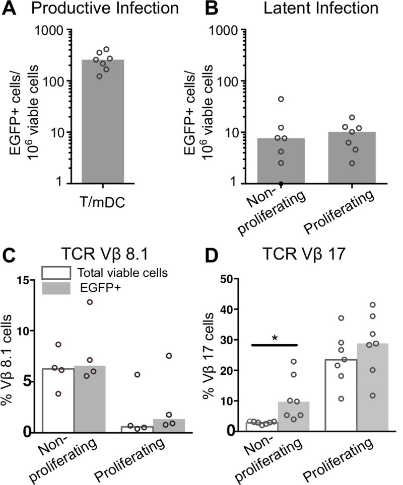
Figure 6. Vβ expression in non-proliferating and proliferating latently infected CD4+ T-cells.
Resting CD4+ T-cells labelled with eFlour670 were cultured for 24 hours with mDC and infected with an EGFP reporter virus. A. Productive infection was measured at day 5 (n=7). At day 5 post-infection, cultures were sorted into non-productively infected, non-proliferating and proliferating CD4+ T-cells. B. Sorted non-proliferating and proliferating T-cells were stimulated using anti-CD3/CD28+integrase inhibitor for 72 hours, to measure latent infection (n=7), and stained with antibodies for C. weak SEB TCR Vβ-8.1 interactions (n=4) and D. strong SEB TCR Vβ-17 interaction (n=7). Columns represent the median in total (clear) or EGFP expressing T-cells (grey) and symbols represent results for individual donors. Significance was measured by paired students T-test where n<5 or Wilcoxon signed-rank test where n≥5, *p≤0.05, **p≤0.005, ***p≤0.0005.

