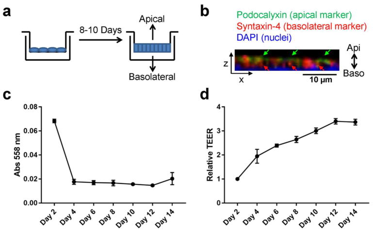
Fig. 1. Polarization of mammary epithelial cells.
(a) Transwell system for the polarization of mammary epithelial cells and collection of apical and basolateral EVs. (b) Immunofluorescence assay of polarized MCF10A cells showing polarized expression of Podocalyxin (an apical marker) and Syntaxin-4 (a basolateral marker). DAPI staining indicates the nuclei. Z-stack images are reconstructed in 3D and the XZ-view is shown. Green and red arrows indicate apical staining of Podocalyxin and basolateral staining of Syntaxin-4, respectively. (c) Time course for diffusion of phenol red from the upper chamber to the lower chamber of MCF10A transwell culture. (d) Time course of trans-epithelial electrical resistance (TEER) from MCF10A cells grown on 10-cm transwell filters.

