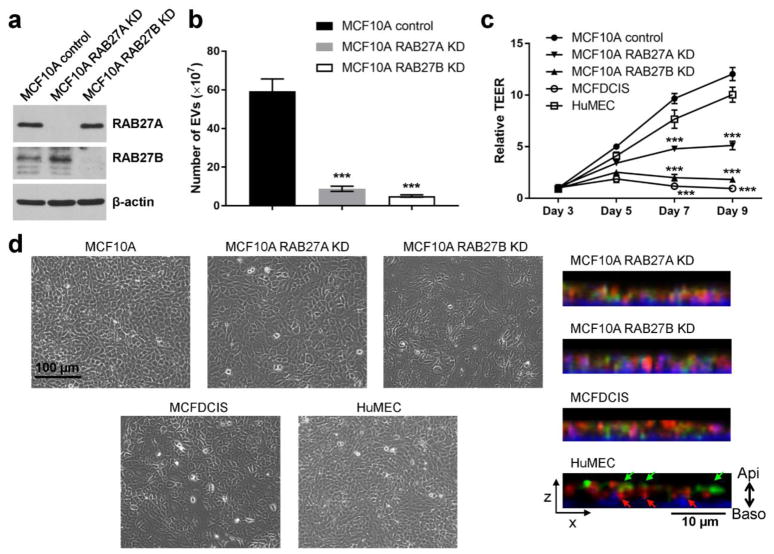
Fig. 3. EV secretion is required for the establishment of epithelial polarity.
(a) Western blot showing indicated protein levels in MCF10A control and RAB27 KD cells. (b) Numbers of EVs secreted by equal number of indicated cells were determined by nanoparticle tracking analysis. *** P < 0.001 compared to the MCF10A control cells. (c) MCF10A (control or RAB27 KD), MCFDCIS, and HuMEC cells were seeded on 24-well transwell inserts and TEER was measured over the indicated time course. *** P < 0.001 compared to the MCF10A control cells. (d) Left: Phase contrast images of indicated cells grown on tissue culture dishes. Right: The XZ-view of reconstructed Z-stack images showing the immunofluorescence staining of Podocalyxin (green) and Syntaxin-4 (red) in cells grown on filters. DAPI staining (blue) indicates the nuclei. Green and red arrows indicate apical staining of Podocalyxin and basolateral staining of Syntaxin-4, respectively.

