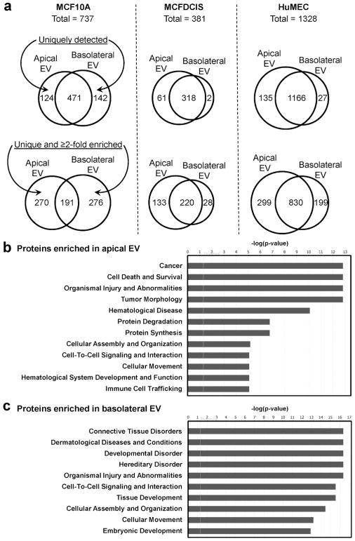
Fig. 5. Identification of proteins in apical and basolateral EVs by mass spectrometry.
(a) Venn diagrams showing the numbers of proteins with at least 4 peptide matches in the apical and basolateral EVs from the MCF10A, MCFDCIS (which shows impaired polarity), and HuMEC models. Numbers of proteins that are uniquely detected from one sample (top), or also including whose enriched by at least 2 folds (bottom) are shown. (b, c) EV proteins identified by mass spectrometry with at least 4 peptide matches and enriched by at least 2 folds in apical (b) or basolateral (c) EVs from both MCF10A and HuMEC models were analyzed by Ingenuity Pathway Analysis for the prediction of associated diseases and functions.

