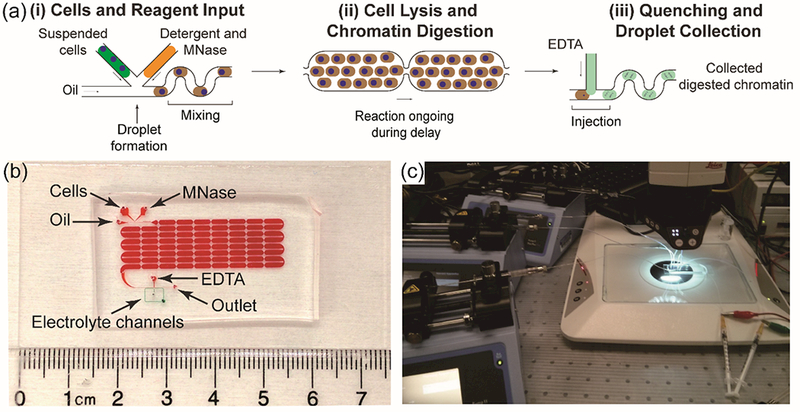
Figure 1.

(a) Schematic of the automated, droplet-based microfluidic nucleosome preparation process. The whole procedure contains three steps: (i) loading cells, detergent, and MNase to the device for droplet generation, (ii) droplets traveling down the delay channel for chromatin digestion to complete, (iii) injecting EDTA solution to droplets to quench the enzymatic processing and collect products. (b) The 8-row device. Delay line and electrolyte channels are highlighted with red and green food dyes, respectively. (c) The complete setup for droplet microfluidic nucleosome preparation.
