Figure 1.
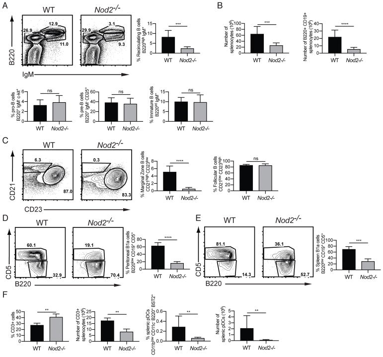
Nod2−/− mice have defects in recirculating B cells, marginal zone B cells, B1a cells. (A) Representative FACS plots and percentage of recirculating B cells (B220high IgM+) and pro-B, and pre-B, and immature B cells in the bone marrow. (B) Number of total live cells, number of B220+ CD19+ B cells, and ratio of immature to mature B cells in the spleen. (C) Representative plots and percentages of marginal zone (CD21 high CD23 low) and follicular (CD21 low CD23 high) B cells in the spleen. Populations are gated on B220+ CD19+ AA4.1-mature B cells. (D–E) Representative plots and percentages of B1a cells (B220 low, CD19+ CD5+) from peritoneal lavage (D) and spleen (E). (F) Percentage and numbers of CD3+ T cells and plasmacytoid dendritic (CD11blow CD11c− B220+ BST2+) cells in the spleen. For A–E, data is from is representative of >5 independent experiments from a total of WT (n = 9) and Nod2−/− (n = 9) mice. For F, data is representative of two independent experiments from a total of WT (n = 4) and Nod2−/− (n = 8) mice. ns p > 0.05, ***p ≤ 0.001, and ****p ≤ 0.0001 by two-tailed Mann-Whitney.

