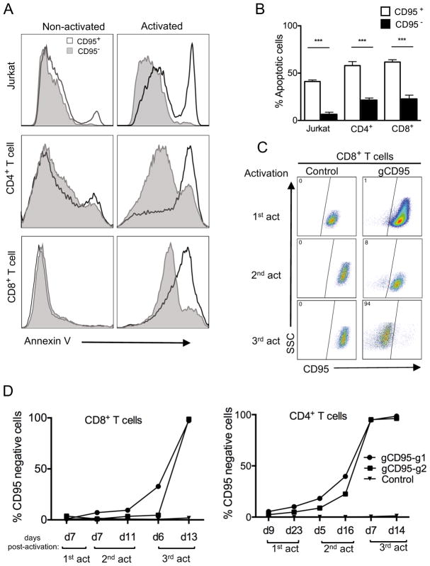
FIGURE 3. Reduced apoptosis in CD95-deleted Jurkat cells and primary human T cell subsets.
(A) Analysis of CD95-induced cell death in CD95-deleted cells. Histogram overlays of Annexin V staining of CD95+ and CD95− cells in activated versus non-activated Jurkat cells and primary CD4+ and CD8+ T cells. Jurkat or primary T cells were activated and transduced with lentivirus targeting the CD95 gene (gCD95) and primary T cells were expanded in IL-2, without selection, then reactivated with anti-CD95 crosslinking antibody or anti-CD3/anti-CD28, respectively, for 24 hours. Reactivated cells were analyzed for apoptosis by Annexin V staining. (B) T cell apoptosis after CD95 gene knockout. Cells were gated on either CD95− or CD95+ 24 hours after reactivation as described in A.Data represent three independent experiments. Error bars represent SEM. ***p<0.001. (C) Preferential survival and expansion of CD95 positive population in CD8+ T cells. CD8+ T cells were transduced and reactivated as in A, through three subsequent restimulations and compared to empty vector control transduced cells. (D) Increased frequency of CD95 negative cells in CD8+ and CD4+ T cell after restimulations. Purified CD4+ or CD8+ T cell subsets were transduced with lentiviruses targeting the CD95 gene using two different gRNA clones (gCD95-g1 or gCD95-g2) and stained for CD95 expression at different time points after three re-stimulations using anti-CD3/anti-CD28 beads. Data is representative of two independent experiments.

