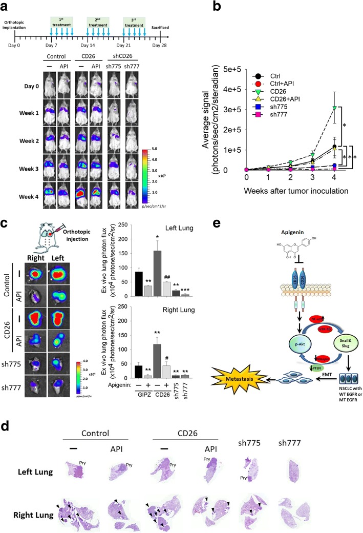
Fig. 6.
Targeting CD26 suppresses non-small cell lung cancer (NSCLC) growth and metastasis by apigenin (API) in an orthotopic mouse model. a, upper panel, Timeline of the in vivo study design for investigating the effects of CD26 expression on tumor progression and the antitumor activity of API. Male NOD/SCID mice were orthotopically injected with luciferase-tagged and CD26-overexpressing or CD26-depleted (sh-775 and sh-777) A549 cells. After 7 days, mice were treated with API (3 mg/kg, IP) or the vehicle for 5 days/week. a, lower panel, All mice were sacrificed at 4 weeks after API treatment, and the luciferase activity was detected every week with a noninvasive imaging system (IVIS imaging system, Xenogen). b Quantitative analysis of Xenogen imaging signal intensity (photons/s/cm2/steradian) every week. * p < 0.05. c Lungs were isolated and examined at the end of this spontaneous metastasis assay. Left panel: Cancer metastasis from the left lung to the right lung was imaged with bioluminescence at the end of the study. Right panel: Signal intensities from primary tumors (left lung) and metastatic tumors (right lung) were bioluminescently captured at the end of the study, with the mean signal for each group indicated. * p < 0.05, ** p < 0.01, *** p < 0.001 compared to the control group. # p < 0.05, ## p < 0.01, compared to the API-treated control group. d H&E staining of lungs isolated from each group of mice; metastatic colonies are indicated by an arrow in the right lungs. e A working model shows the molecular mechanism underlying the ability of API to suppress the metastasis of NSCLC cells. The antimetastatic activity of API on NSCLC cells with wild-type or mutant epidermal growth factor receptor (EGFR) was attributed to inhibition of CD26 expression following suppression of the interplay between Akt activation and Snail/Slug expression, with ultimate restraint of EMT progression. Red ovals indicate hypothetical regulators which might be involved in the API-mediated interplay of Akt and Snail family members

