Fig. 6.
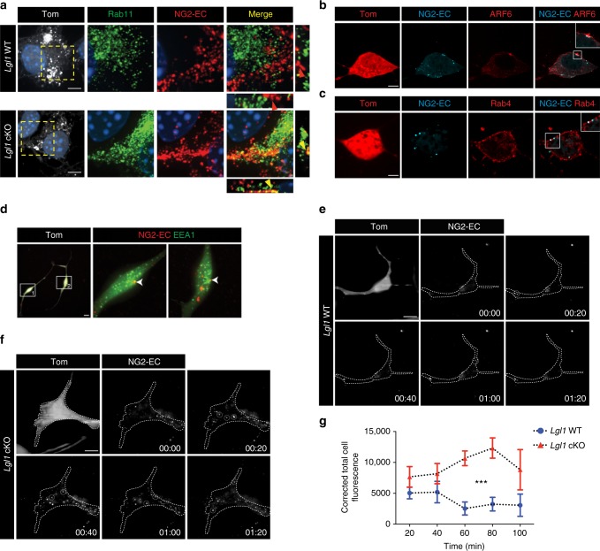
Non-degraded NG2 in Lgl1 cKO OPC is recycled to the membrane. a Representative co-immunostaining for NG2-EC (red) and Rab11 (green) 45 min after incubation in differentiation medium. Note the higher colocalization of NG2-EC with Rab11 in Lgl1 cKO OPC (yellow arrowheads) compared to Lgl1 WT cells (red arrowhead). Scale bar: 5 μm. b–d Fluorescent immunostaining pictures illustrating colocalization of remaining NG2 (blue) with (b) ARF6 (red), (c) Rab4 (red), and (d) EEA1 (green) in Lgl1 cKO cells 24 hours after incubation in differentiation conditions. Scale bars: 10 μm. e, f Frames illustrating TIRF time lapse of Lgl1 cKO and WT cells 4 hours after incubation in differentiation conditions. Pictures were taken each 20 min. Examples of recycling labeled NG2 puncta (white) are highlighted in dashed circle. NG2 recycling persists in Lgl1 cKO cells up to time point 01:20 whereas it is insignificant in WT cells. Time indicated as hours:minutes. Scale bar: 10 μm. g Graph representing quantification of the corrected total cell fluorescence reflecting NG2 recycling in Lgl1 cKO cells (blue circles) and in Lgl1 WT cells (red lozenges). Data are depicted as mean ± s.e.m., n = 8 cells for each genotype, three independent experiments (***p < 0.01, two-way ANOVA)

