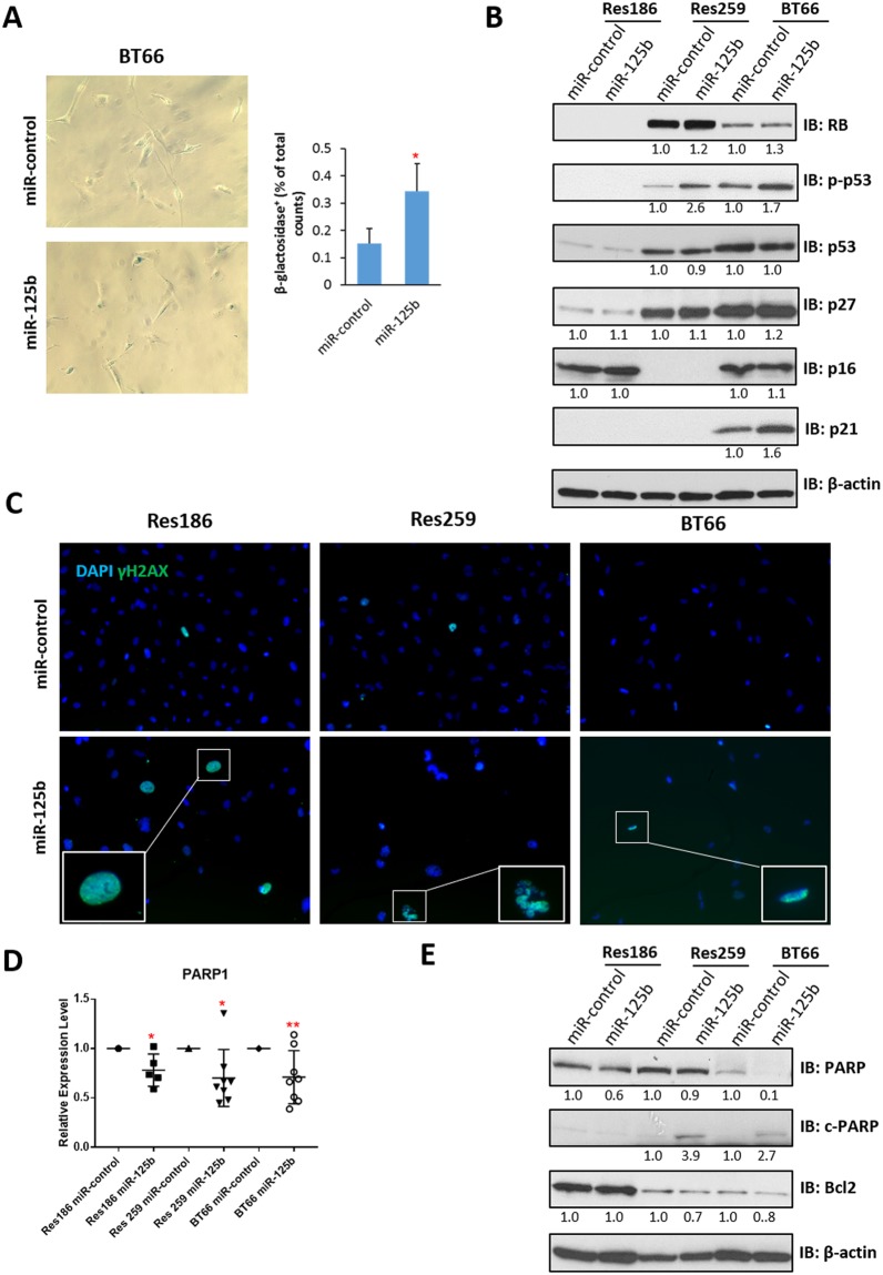
Figure 7.
Overexpression of miR-125b induces senescence and apoptosis. (A) Representative photomicrographs (200x) of β-galactosidase staining on BT66 cells infected with miR-control or miR-125b, quantification of staining is at right. (*p < 0.05 compare to miR-control). (B) Western blot of senescence proteins p16, p21, p27, p53 and RB. β-actin was used as loading control. Protein levels were quantified and normalized to miR-control. (C) Representative photomicrographs (200x) of γ-H2AX shows increasing of γ-H2AX positive cells with miR-12b overexpression in all cell lines tested. (D) qRT-PCR of PARP1 expression in Res186, Res259 and BT66 (BRAF fusion+) cells infected with miR-control or miR-125b. All data were normalized to miR-control for each cell lines. (*p < 0.05, **p < 0.01). (E) Western blot of apoptotic protein PARP and the anti-apoptotic protein Bcl2. β-actin was used as loading control. Protein levels were quantified and normalized to miR-control (uncropped Western blots presented as Supplementary Fig. 4). Results represent the average of at least three biological replicates and data were analyzed with a two-tailed Student’s t-test.

