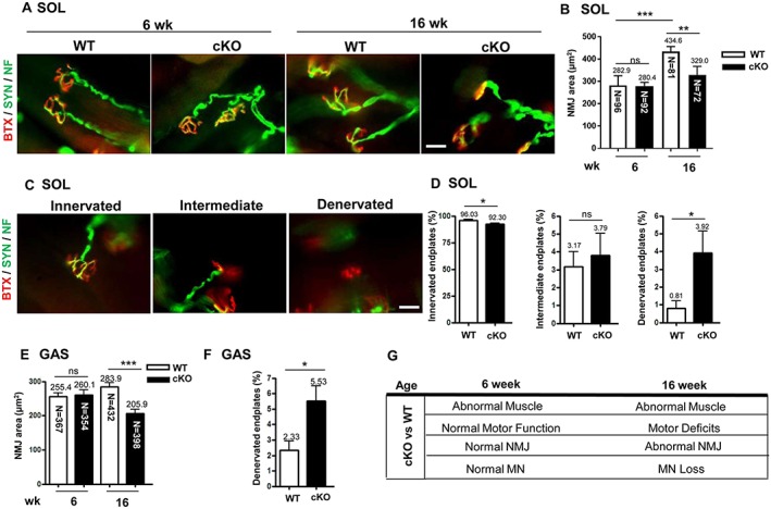
Figure 3.

The abnormal architecture of the neuromuscular junction (NMJ) with axonal denervation in conditional knockout (cKO) mice. (A) Fluorescent images of NMJ in soleus muscle (SOL) from mice at age 6 and 16 weeks. Immunofluorescence assay with α‐bungarotoxin (BTX) [red, for acetylcholine receptor (AChR)] and anti‐synaptophysin (Syn) and anti‐neurofilament (NF) antibodies (both green, for nerve terminals) used for analysis of NMJ area and the patterns of axonal innervation. Scale bar: 10 μm. (B) Quantification of NMJ area in soleus muscle in mice at age 6 and 16 weeks. The area of BTX‐labelled AChR represents the NMJ area. n = 5 for 6 week and n = 7 for 16 week mice. The number in each bar indicates the number of examined NMJs. (C) Patterns of axonal innervation in mice at age 16 weeks. Three NMJ innervation patterns are indicated, including innervated (well overlapping of AChR and nerve terminals), intermediate (nerve terminal adjacent to AChR without overlapping), and denervated (no nerve terminal adjacent or overlapped to AChR). n = 3 for each group. Scale bar: 10 μm. (D) Proportion of innervated, intermediate, and denervated endplates in soleus muscle from mice at age 16 weeks. (E) Quantification of NMJ area in gastrocnemius muscle (GAS) from mice at age 16 weeks and (F) proportion of denervated endplates in gastrocnemius muscle. n = 3 for each group. (G) Pathological and motor functional results in cKO mice at age 6 and 16 weeks. Data are mean ± SEM. Panels D and F: by Student's t‐test; panels B and E: one‐way analysis of variance. * P < 0.05, ** P < 0.01, and *** P < 0.001. ns, not significant; WT, wild‐type.
