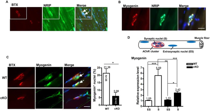
Figure 5.

Low myogenin expression at the neuromuscular junction (NMJ) in conditional knockout (cKO) mice at age 16 weeks. (A) Immunofluorescence assay with anti‐nuclear receptor interaction protein (NRIP) antibody (green), bungarotoxin (BTX) (red), and DAPI (blue) of NRIP localized around acetylcholine receptor (AChR) in longitudinal sections of soleus of wild‐type (WT) mice (arrows indicate co‐localization of NRIP and AChR), (B) co‐localization of NRIP (green) and myogenin (red) at NMJ with clustered nuclei (DAPI, blue) in soleus muscle WT mice, and (C) proportion of myogenin (green)‐immunoactive AChR (red) in soleus muscle of cKO and WT mice (arrows indicate co‐localization of myogenin and AChR cluster). Scale bars, 100 μm. The number in each bar indicates the number of examined NMJs. n = 3 for each group. (D) Gastrocnemius muscles of mice at age 16 weeks subjected to laser capture microdissection. RNA was extracted from extrasynaptic and synaptic regions, and the amount of myogenin transcript was analysed by real‐time quantitative PCR. Data were normalized to β‐actin level. n = 7 for each group. Data are mean ± SEM. Panel B: by Student's t‐test; panel D: one‐way analysis of variance. * P < 0.05, ** P < 0.01, and *** P < 0.001.
