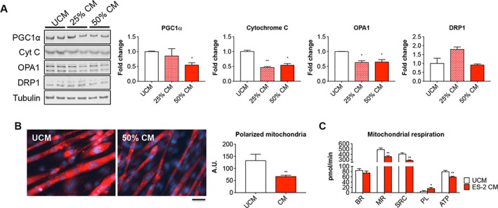
Figure 11.

Mitochondrial homoeostasis and respiration are impaired in C2C12 myotubes exposed to ES‐2 conditioned medium (CM). Representative western blotting and quantification for PGC1α, Cytochrome C (Cyt C), OPA1, and DRP1 in protein extracts from C2C12 myotubes exposed to different concentrations of ES‐2 CM (n = 3). Tubulin was used as loading control (A). Mitochondrial polarization assessed by means of MitoTracker red CMXRos staining in C2C12 exposed to 50% ES‐2 CM (n = 3). Red signal intensity was quantified by using the ImageJ software (five fields per sample). Scale bar: 100 μm (B). Basal respiration (BR), mitochondrial respiration (MR), spare respiratory capacity (SRC), proton leak (PL), and ATP production (data expressed in pmol/min) determined by extracellular flux analysis in C2C12 myotubes exposed to 50% ES‐2 CM (n = 3) (C). Data are reported as means ± standard deviation. Significance of the differences: * P < 0.05, ** P < 0.01, *** P < 0.001 vs. unconditioned medium (UCM).
