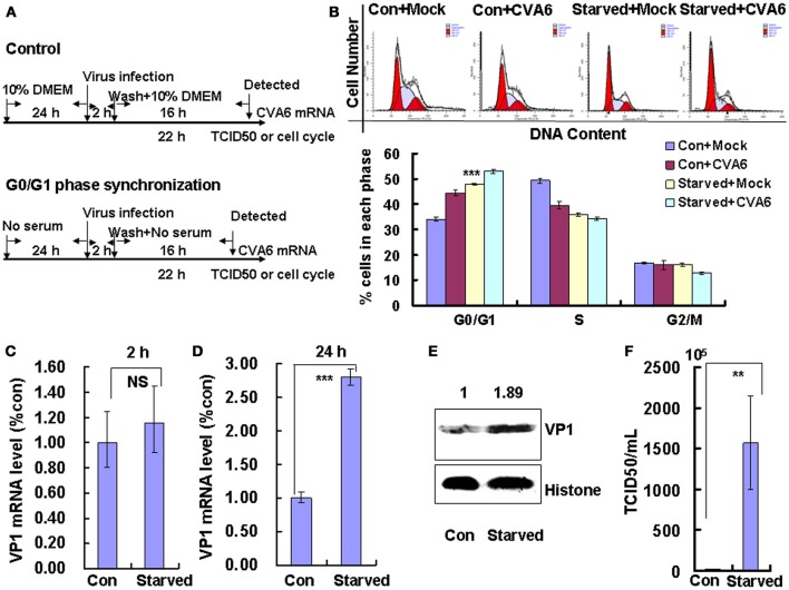
Figure 2.
G0/G1 phase-synchronization promotes viral replication. (A) RD cells were cultured in serum-free medium for 24 h for G0/G1-phase synchronization. Infected with mock (Mock) or infected with CVA6 (CVA6) at an MOI of 1 for 2 h, then the medium was restored to maintain the cell cycle synchronization status for 24 h. (B) Top panel: Flow cytometry determined the cell cycle profiles after culture in control medium (Con) or serum-free medium (Starved) and mock-infection or infection with CVA6. Bottom panel: The histograms indicating cell cycle distribution were analyzed by the ModFit LT program. ***P < 0.001 (Starved+Mock vs. Con+Mock). (C,D) Intracellular CVA6 RNA levels were detected by quantitative real-time PCR in RD cells that were cultured in control (Con) or serum-free medium (Starved) at 2 h (C) or 18 h (D) post-infection with CVA6. The results were standardized using GAPDH mRNA as a control and normalized to 1.0 in the Con cells. (E) The expression of VP1 was determined in control medium (Con) or serum-free medium (Starved)-treated cells at 24 h post-infection by Western blot analysis. Histone is the loading control. The results are representative of three independent experiments. (F) The total progeny viruses (the supernatant and intracellular viruses) were titrated in RD cells, and the TCID50/mL value was determined at 24 h post-infection. The results represent the mean ± SD of three independent experiments. NS, no significant difference; **P < 0.01 and ***P < 0.001.

