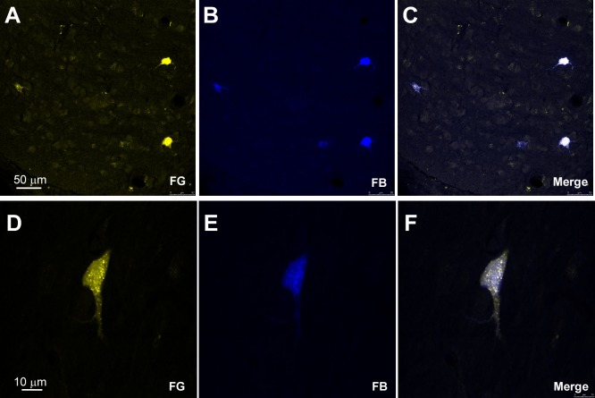
FIGURE 3.
Microphotographs of HDB labeled neurons after injections/deposits in S1/V1. (A–C) Confocal images showing single and double-labeled neurons in HDB in a representative animal; the merge shows the presence of double-labeled neurons. (D–F) Another examples of double-labeled neurons at high magnification.

