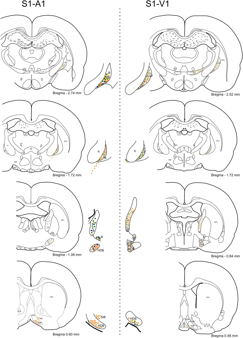
FIGURE 5.
Rostral to caudal schematic drawings of brain coronal sections of BF neurons projecting to S1, A1, and V1 cortices. Neurons projecting to S1 or/and A1 cortices are shown on the left column (o similar) of the figure and to S1 or/and V1 cortices on the right column of the figure. Neurons are depicted as: single-FG (orange, injection in S1), single-FB (blue; injection in A1), single-FB (blue; injection in V1), and double-labeled neurons (green). Notice the different location in the BF nuclei of neurons projecting to S1, A1, or V1 cortices. 3V, 3rd ventricle; aca, anterior commissure; CPu, caudate putamen; EGP, external globus pallidus; Re, reuniens thalamic nucleus; SIB, substantia innominate basal part; SO, supraoptic nucleus; VCl, ventral part of claustrum.

