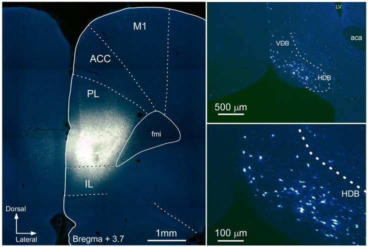
FIGURE 8.
Microphotograph of a coronal section at Bregma +3.72 mm showing the location of the FG injection in PL/IL cortices (Left). Microphotographs at different magnifications of FG labeled neurons in HDB (Right). ACC, anterior cingulate cortex; fmi, corpus callosum forceps; LV, lateral ventricle; M1, primary motor cortex; rest of lettering as in Figure 5.

