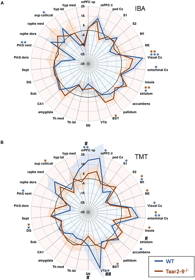
FIGURE 8.
Region-specific neural activity of Taar2-9−/− and wild type (WT) mice stimulated with isobutylamine (IBA) and trimethylthiazoline (TMT). Neural activity was assessed by functional magnetic resonance imaging (fMRI) based on continuous arterial spin labeling (CASL) which reports cerebral perfusion as a proxy for neural activity. Represented are means (solid lines) ± SEM (shaded area) of region-specific normalized perfusions elicited by (A) IBA and (B) TMT of 7 to 12 Taar2-9−/− males and WT littermates. Blue ∗ and red ∗ denote statistically significant (∗P < 0.05, ∗∗P < 0.01, ∗∗∗P < 0.001) differences between odor exposed and non-exposed Taar2-9−/− and WT mice, respectively. #Denotes statistically significant (#P < 0.05, ##P < 0.01) differences between Taar2-9−/− and WT mice. mPFC vp, ventral prelimbic medial prefrontal cortex; mPFC il, ventral infralimbic medial prefrontal cortex; ped Cx, dorsal peduncular cortex; S1, primary somatosensory cortex; S2, secondary somatosensory cortex; M1, primary motor cortex; M2, secondary motor cortex; Cx, cortex; BST, bed nucleus of the stria terminalis; VTA, ventral tegmental area; SN, substantia nigra; Th lat, lateral thalamus; Th med, Medial thalamus; CA1, hippocampal area CA1; Sub, subiculum; DG, dentate gyrus; Sept, septum; PAG dors, dorsal periaqueductal gray; PAG vent, ventral periaqueductal gray; raphe dors, dorsal raphe; raphe med, median raphe; sup colliculi, superior colliculus; hyp lat, lateral hypothalamus; hyp med, medial hypothalamus.

