
FIGURE 1.
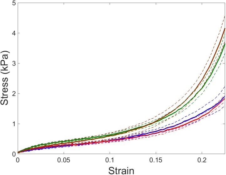
Stress–strain relationships of left ventricular (LV) myocardium ECM. Stress–strain recorded by tensile stretching in ECM myocardium strips of young and aged mice subjected to room air (blue and red lines, respectively) and intermittent hypoxia (IH) exposures mimicking obstructive sleep apnea (OSA) (green and brown lines, respectively). Data are mean (solid lines) ± SE (dashed lines).