Figure 1.
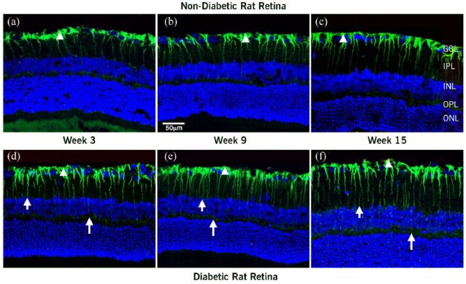
Confocal images of localization of Ang II and nuclei merge in retina sections at 3, 9, and 15 weeks after diabetes was induced: (a–c) nondiabetic and (d–f) diabetic rats. In the nondiabetic rats, Ang II antiserum labeled Müller cells. Ang II extended from the footplates of the Müller cell (arrowhead) through cell processes in the IPL into the INL. Immunoreactivity was marked in the Müller cell endfeet and extended into the cellular processes. In the diabetic retina, Ang II extended through the entire retina from the Müller cell footplates (arrowhead), cell processes in the IPL through the INL and OPL into the ONL (short and long arrow). The higher intensity and increase in extent of labeling in Müller cell processes was clear at three weeks after diabetes was induced. This pattern of labeling was maintained throughout the 15 weeks of diabetes.
GCL: ganglion cell layer; IPL and OPL: inner and outer plexiform layers; INL and ONL: inner and outer nuclear layers.
Scale bar = 50 µm.

