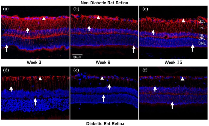
Figure 2.
Confocal images of localization of Ang-(1-7) in retinal sections. Confocal images of Ang-(I-7) and nuclei merge in retina at 3, 9, and 15 weeks after diabetes was induced: (a–c) nondiabetic and (d–f) diabetic rats. Ang (1-7) antiserum-labeled Müller cells (arrowhead). In the nondiabetic retina, Ang-(I-7) extended from the footplates of the Müller cell (short arrow) through cell processes extending up to the photoreceptor layer (long arrow). In diabetic rats, the intensity and extent of labeling decreased steadily with the duration of hyperglycemia.
Scale bar = 50 µm.

