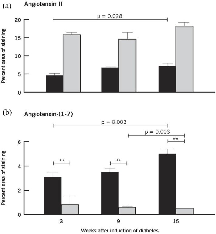
Figure 3.
Quantitative evaluation of Ang II (a) and Ang-(1-7) (b) in nondiabetic and diabetic retinas at 3, 9, and 15 weeks after induction of diabetes. Values are mean ± SEM. Within group: Ang II and Ang (1-7) showed differences among the three time points only in the nondiabetic retinas (p = 0.028 and p = 0.003, respectively). Mean levels of Ang II in the diabetic group were statistically significantly higher than those in the nondiabetic group (**p < 0.001). Mean levels of Ang-(1-7) in the diabetic group were statistically significantly lower than those in the nondiabetic group (**p < 0.001). Nondiabetic retina (black square); diabetic retina (gray square).

