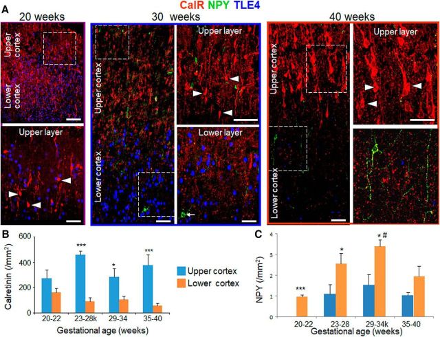
Figure 3.
CalR is enriched in upper cortical layer and NPY is enriched in the lower cortical layer. A, Representative immunofluorescence of cryosections from upper and lower cortical layers of 20, 30, and 40 gw infant (as indicated) labeled with CalR-, NPY-, and TLE4-specific antibodies. For the 20 gw image, bottom shows high-magnification of the boxed area at the top. For the 30 and 40 gw infant, images on the right are high-power views of the boxed areas on the left. Note higher density of CalR in the upper cortical layer compared with lower cortical layer. Note relatively fewer number of NPY+ cells compared with the density of CalR. Scale bar, 50 μm for the left and 20 μm for the right panel for each image. B, Data are shown as mean ± SEM (n = 5 each group). The density of CalR+ cells was consistently higher in upper compared with lower cortical layer for 23–28, 29–34, and 35–40 gw infants. *p < 0.05 and ***p < 0.001 for comparison between upper versus lower cortical layer within a gestational age category. C, Data are shown as mean ± SEM (n = 5 each group). NPY+ interneurons in the lower cortical layer were higher in density in 29–34 gw compared with 20–22 gw infants. NPY+ interneurons were more abundant in lower compared with upper cortical layer as indicated. *p < 0.01, ***p < 0.001 for the comparison between upper and lower cortical layer within a gestational age category; #p < 0.05 for the comparison between 20 and 22 and 29–34 gw within lower cortical layer.

