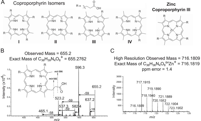
FIG 3 .
Mass spectrometry for identification of coproporphyrin. Tandem mass spectrometry strongly suggested a coproporphyrin, and high-resolution mass spectrometry matched zinc coproporphyrin. (A) Structural isomers of coproporphyrin are distinguished by the orientation of R groups around the porphyrin ring. (B) MS/MS fragmentation patterns of m/z 655 suggested a coproporphyrin identification but did not distinguish between structural isomers I to IV. Retention time matching was used to confirm identity of the coproporphyrin III isomer (Fig. 4). (C) High-resolution analysis of m/z 716 showed isotopic patterns consistent with that of zinc coproporphyrin III within acceptable parts-per-million error range.

