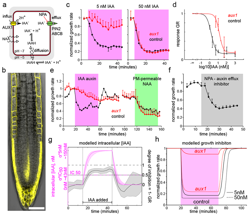
Fig.3.

The rapid root growth inhibition depends on auxin levels inside the cell. (A). A schematics of auxin fluxes in a root epidermal cell. (B). Auxin influx carrier AUX1-YFP is expressed in the root epidermal cells. Scale bar = 50µm. (C). The aux1 mutant is resistant to 5nM IAA but reacts to 50nM IAA. Mean normalized values of 3 (5nM), 2 (50nM control) and 5 (50nM aux1) roots, ±SD. (D). The dose response of aux1 is shifted towards higher IAA concentrations by ~10 fold (control is the same as in Fig.1E). Fit: IC50aux1=17.2nM = 11.8*IC50col . Data points are means of N = 10,4,2,9,7 roots for [IAA]ext =5,10,20,50,1000nM respectively +-SD; the 1000nM was done on agar plate. (E). The aux1 mutant reacts little to 5nM IAA (magenta) but its reaction to membrane permeable NAA (100nM, green) is comparable to control roots. Mean normalized values of 5 (aux1) and 2 (control) roots, ±SD. (F). The auxin efflux inhibitor NPA (5µM, gray) triggers root growth inhibition. Mean normalized values of 5 roots, +SD. (G). Auxin accumulation (magenta, simulated) is faster than growth inhibition during application of 5nM IAA (gray, mean experimental values of 1-GR). Note time delay between [IAA]cell = αcol · IC50 and (1 – GR) = 0.5. Accumulation ratio αcol ≅ 30 (Supp.text). Shaded regions correspond to 1 SD (gray, n=6 roots) or variation due to SD of parameter values (magenta, simulation results). (H). The modelled theoretical inhibition for aux1 and control roots.
