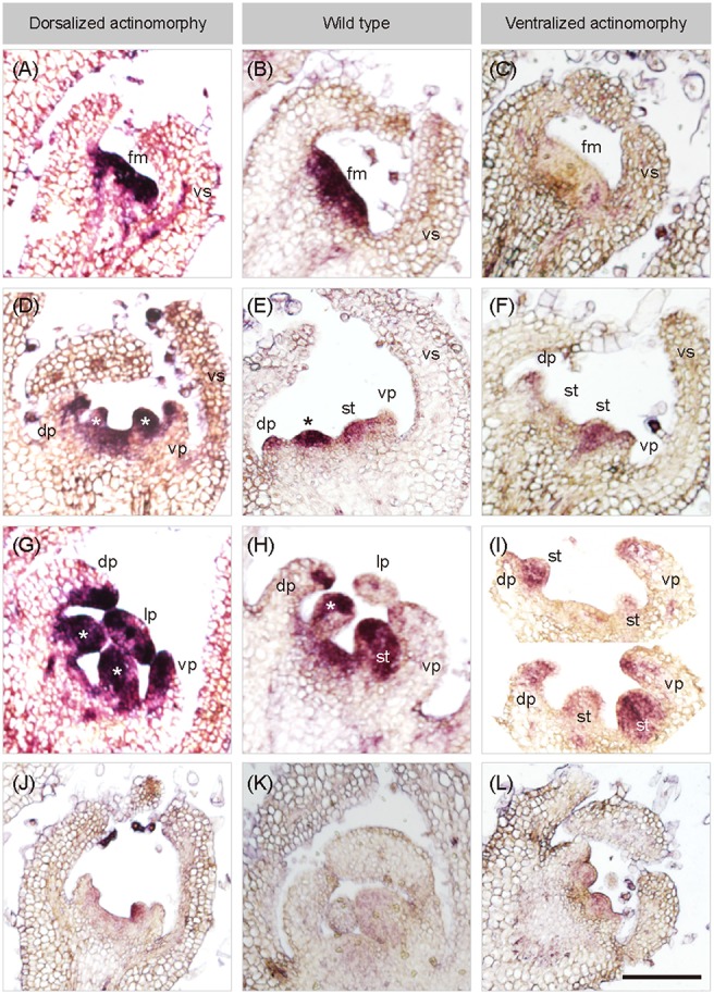
Figure 4.

In situ hybridization of SiCYC1A in flower buds of African violet. Longitudinal sections on early flowering stages of all cultivars hybridized with antisense probes of SiCYC1A: stage 4 (A–C), stage 5 (D–F) and stage 6 (G–I). The purple color represents the in-situ signals of SiCYC1A mRNA. SiCYC1A has early expression in the entire floral meristem of DA and WT (A,B) but is absent in VA (C). During floral organ initiation (D–F), SiCYC1A persists in inner whorls of the flower including the dorsal (dp) and ventral petal primordia (vp) of DA (D). (E) But in zygomorphic WT, SiCYC1A confines more to the dorsal petal primordia (dp) than the ventral one. (F) In VA, weak SiCYC1A signals start to acuminate at tips of both dorsal and ventral petal primordia. (G) Later in floral organ differentiation, SiCYC1A can be detected in all petals (dp, lp and vp) of DA. (H) But in WT, SiCYC1A is largely restricted to dorsal petals (dp). (I) In the petal whorl of VA, a weak but slightly stronger SiCYC1A is evident in dorsal petals (dp) when compared to ventral petals (vp). The sense probe controls were shown in (J–L). The cross sections of ISH are shown in Figure S5. fm, floral meristem; vs, ventral sepal; dp, dorsal petal; lp, lateral petal; vp, ventral petal; st, stamen. *, staminode. Scale bar = 100 μm.
