Figure 1.
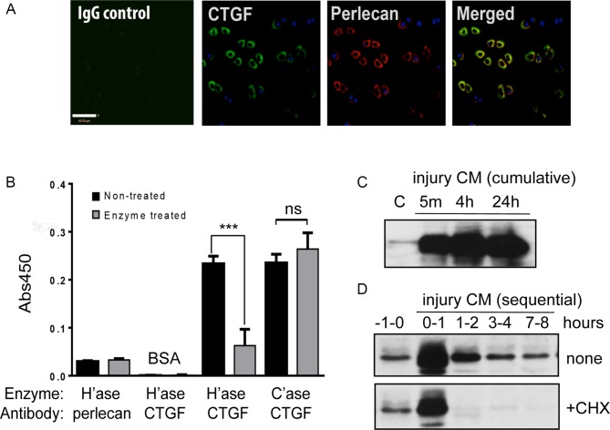
Connective tissue growth factor (CTGF) binds to perlecan in the pericellular matrix of articular cartilage and is released rapidly on injury. (A) Confocal microscopy of normal human articular cartilage, showing pericellular colocalisation of CTGF (green) and perlecan (red). Scale bar, 40 µm. (B) bovine serum albumin (BSA) or perlecan was precoated onto ELISA plates and wells were treated with or without 10 mU/mL of heparitinase (H’ase) or chondroitinase (C’ase) prior to incubation with 0.05 µg recombinant CTGF for 3 hours. CTGF was detected with anti-CTGF antibodies using a standard ELISA plate reader. Levels of bound perlecan pre-enzyme and post-enzyme treatment were checked with an anti-perlecan antibody. (C, D) Porcine articular cartilage explants (5×4 mm discs) were rested in serum-free (SF) medium for 48 hours and re-cut in fresh SF medium in the presence or absence of 10 µg/mL cycloheximide (+CHX). Injury conditioned medium (CM) was collected cumulatively (C) or sequentially (D) after specific time points and immunoblotted for CTGF. ***P<0.001; ns, not significant by a two-sided Student’s t-test.

