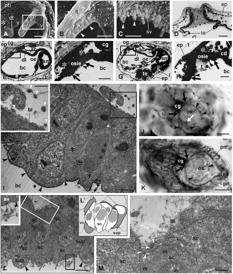
Fig. 2.
Coronal organ in adult blastozooids and differentiation in early buds. (A–D) Adult. (A–C) SEM of part of the oral siphon viewed from the branchial chamber to show the coronal organ. The square area in A is enlarged in B. Arrowheads point to cilia of coronal sensory cells. Asterisk: ciliated area close to the dorsal tentacle (dt). (D) Transverse section of an oral siphon; the tunic (t) lies on its inner side, partly covering the velum and tentacle (te) upper side. The section is tangential to the siphon wall so that the tunic appears to fill the siphon lumen. Toluidine blue. (E–H) Sagittal medial sections of two early cycle buds at the beginning of the cycle (earlier stage in E–F). The square areas in E and G are enlarged in F and H, respectively, to show the details of the oral siphon rudiment. The oral siphon inner epithelium (osie, borders marked by arrowheads) and epidermis are covered by the tunic; asterisks: muscle cell precursors. Arrows: velum area. Toluidine blue. (I) Mitotic cells (asterisks) within the velum/tentacles epithelium. Black arrowheads: thin tunic layer covering the apex of the oral siphon inner epithelium. The squared area is enlarged in the inset. White arrowheads: neurites close to the velum/tentacles area. TEM. (J–K) AChE reaction on a bud in early- (J) and mid-cycle (K) in dorsal view. Arrowheads: neurites reaching the oral siphon rudiment. The cerebral ganglion and nerves are marked. White arrow in J: forming right lateral tentacle marked by the AChE reaction. Black arrows: anterior nerve roots; pn, pericoronal nerves. Anterior at right-bottom. Whole mount bud. (L–M) Detail of differentiating coronal cells in a mid-cycle bud. Oblique cut of the organ in L shows a few sensory cells (schematized in LI; white cells: sensory cells; gray cells: supporting cells; dotted cells: velar cells). Black square, inset in L: stereovilli (sv) and a cilium (c) of a hair bundle, both of which are covered by the fibrillary fuzzy coat. Black arrowheads: thick glycocalyx covers supporting (sup) and velar (vc) cells. Sensory cells lack the covering. In M, the basal groove is enlarged with neurites (white square in L). White arrowheads: first recognizable synaptic contacts. ac, atrial chamber; as, atrial siphon; bc, branchial chamber; bl, basal lamina; cd, ciliated duct of the neural gland; cg, cerebral ganglion; dl, dorsal lamina; e, endostyle; ep, epidermis; i, intestine; lt, lateral tentacle; m, mitochondrion; n, nucleus; oe, esophagus; os, oral siphon; pb, peripharyngeal band; sc, sensory cell; sm, stomach; te, testis lobule; v, velum. Scale bars: A, D, and E, 50 µm; B, F, and G, 20 µm; C, I, and L, 2 µm; H, 5 µm; I (inset), 1 µm; J and K, 100 µm; M, 500 nm.

