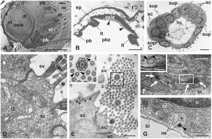
Fig. 3.
Coronal organ differentiation in mid bud. (A) SEM of the oral siphon rudiment viewed from the branchial chamber. The siphon is closed. The peripharyngeal band (pb) marks the border between the anterior prebranchial zone and posterior branchial zone of the pharynx. Developing velum (v) and first order tentacles are visible (lt, the two lateral ones; dt, the dorsal one; vt, the ventral one). Stigmata (st) are perforated. (B) Transverse section of the oral siphon rudiment showing the relationships between the two lateral tentacles (lt), the peripharyngeal band (pb), and the oral siphon rudiment. Arrowheads: tunic produced by the oral siphon inner epithelium (osie). Toluidine blue. (C) Transverse section of a tentacle belonging to a bud at a more advanced stage than that shown in A–B. Note the slightly spoon-shaped upper surface facing the inflowing seawater (upper, left). hc, hemocytes in the blood sinus. TEM. (D–E) Details of the sensory bundle, cut longitudinally in D, transversely in E. Stereovilli and cilium are linked to one another by the fibrillary fuzzy coat (asterisks in D, black arrowheads in inset of E). White arrowheads in D: microfilaments in stereovilli. TEM. G, Golgi field. (F–G) Details of sensory cell basal grooves containing neurites (ne) separated from the hemocoel by the basal lamina (bl). Some synaptic contacts are recognizable (white arrows in F). Some vescicles (arrowheads) are within neurites (enlarged in inset in F). TEM. Other symbols as in Fig. 2. Scale bars: A, 100 µm; B, 50 µm; C, 3 µm; D and F, 300 nm; inset in F: 150; and E and G, 600 nm.

