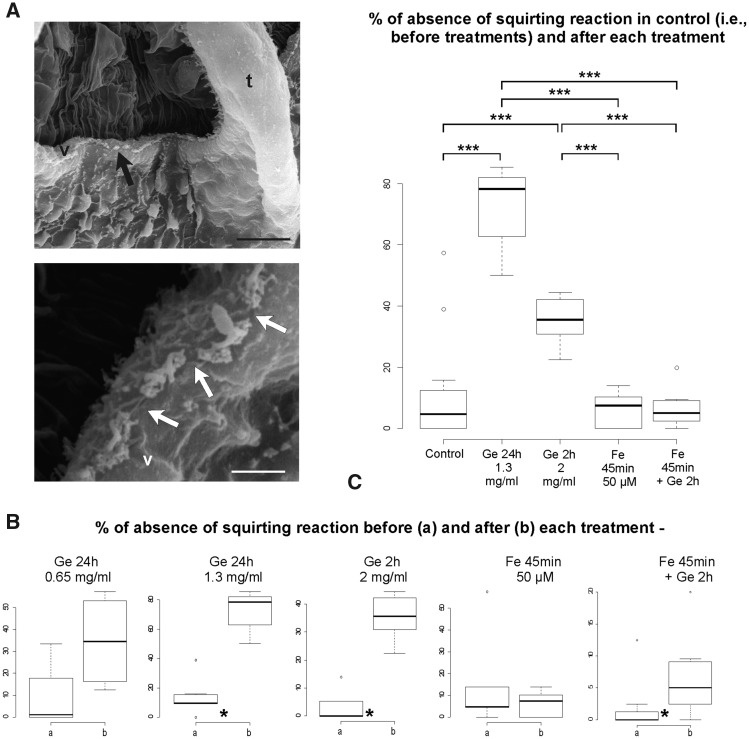
Fig. 4.
Gentamicin-induced morphological and mechanosensorial impairments on coronal organ. (A) Colonies treated with gentamicin. SEM of part of two oral siphons viewed from the branchial chamber to show the coronal organ on the velum (v) and tentacles (t). Arrows point to some discontinuity of coronal apical tufts. (B–C) Boxplots of datasets for the following: (B) comparison of means for two samples of paired data (before and after each single treatment) and (C) ANOVA and post hoc test among treated samples (samples obtained from data after each treatment) and control (a sample obtained from all data before treatment). Boxplots from the datasets obtained from null plus faint reactions are shown. Ge, gentamicin; Fe, fenofibrate. Asterisks denote significance in the comparisons between samples as follow: * <0.05, ** <0.01, *** <0.001. Scale bars in A: 10 µm (upper) and 2 µm (lower).

