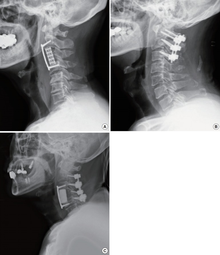
Fig. 2.

Postoperative radiographs presenting each approach, which was determined according to the tumor location, level of the cervical spine, and number of levels involved. (A) An anterior approach was performed with a mesh cage filled with autograft bone and an anterior plating system. (B) A posterior approach was carried out with decompressive laminectomy and a lateral mass and pedicle screw-rod system. (C) A combined approach was performed with polymethylmethacrylate to reconstruct the vertebral body, an anterior plating system, and a lateral mass screw-rod system.
