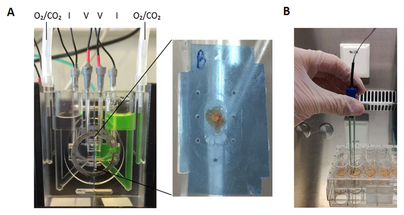
Figure 2. Ex vivo and in vitro methods to assess epithelial barrier function.
(A) Ussing chamber assay: An endosopic intestinal biopsy is placed on one half of the chamber with the mucosal side facing upwards. The tissue is gently placed in-between plastic films for mounting. After equilibration of the tissue for 30 minutes, fluorescently labelled dextrans (FD4) are added to the mucosal side of the chamber. Serosal samples are taken every 30 minutes for 2 hours to evaluate the passage through the biopsy. The transepithelial electrical resistance (TEER) is recorded every 30 minutes by the electrodes. (B) Transwell assay: Intestinal cells are grown on permeable filter supports. Medium is changed every two days after seeding the cells. TEER measurements are performed using chopstick electrodes to measure confluency of the cells.
FD4, FITC-dx4, fluorescein isothiocyanate, dextran 4000 Da; TEER, transepithelial electrical resistance

