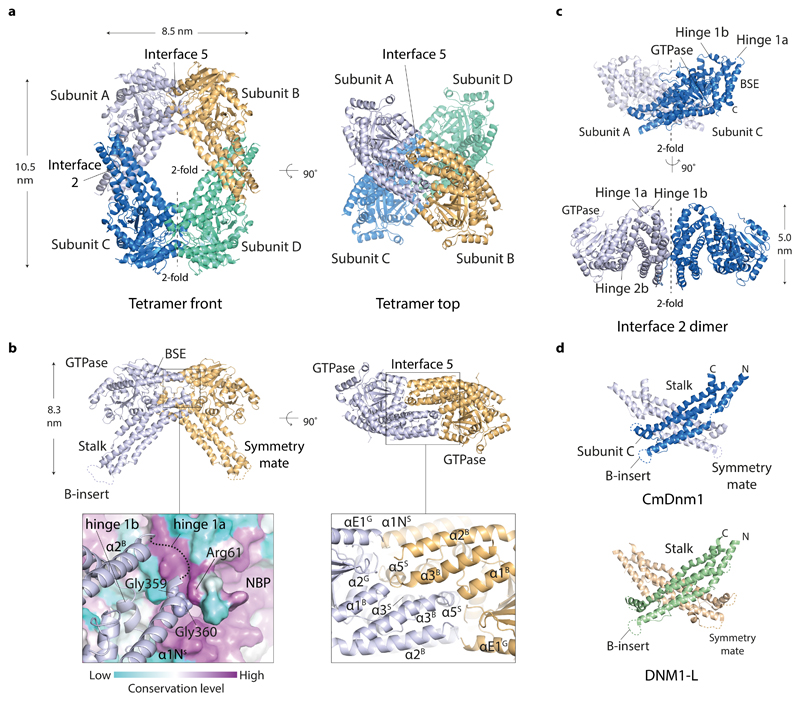
Figure 3. The crystal structure of the CmDnm1 tetramer.
a, CmDnm1 packs as a diamond-shaped tetramer generated by crystallographic 2-fold axes around interface 2 and interface 5. Note how interface 5 dimers are offset by a ~ 90° screw or twist around the tetramer long axis. b, Cartoon schematic showing the arrangement of the interface 5 dimer. (Left) Front view of the interface 5 dimer. Zoom panel shows surface colouring based on residue conservation level calculated against 150 CmDnm1 homologues. Hinge 1a nestles in a highly conserved groove at the backside of the nucleotide binding pocket and contacts Arg61 located next to the Switch 1 Thr60. NBP = nucleotide binding pocket. (Right) Top view of interface 5 dimer. Zoom panel shows key helices contributing to the interface. c, Cartoon schematic showing the highly conserved interface 2 dimer. d, CmDnm1 and DNM1-L (PDB 4BEJ) form equivalent interface 2 stalk dimers.

