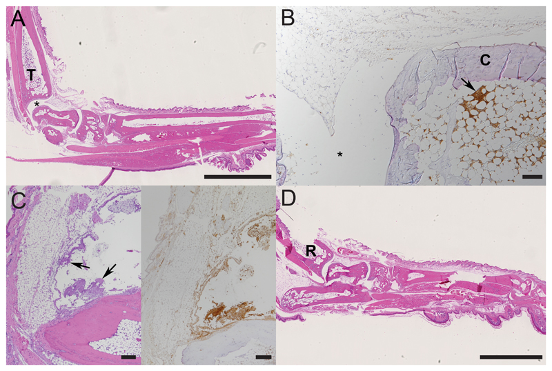
Figure 4. Histological findings in paws with collagen-induced arthritis after one dose of treatment.
A, B. Right hindleg of a mouse 12h post treatment (score 0; initial score 2) with neutrophils only found in bone marrow, T - tibial bone; * joint space; C – cartilage. C. Left hindleg of a mouse 24h post treatment (score 0; initial score 2). D. Right foreleg of a mouse 72h post treatment (score 0; initial score 3), R - radial bone. B, C: Arrows: accumulations of viable and degenerate neutrophils. A, C (left), D: HE stain; B, C (right): staining for neutrophils (Ly6G+); ABC method, hemalaun counterstain. Bars = 2.5 mm (A, D) and 100 μm (B, C).

