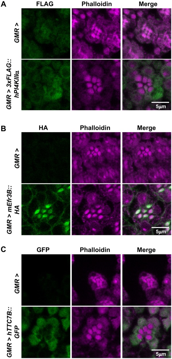
Fig. 6.

Localization of mammalian PI4KIIIα, Efr3 and TTC7 in photoreceptors. (A) Confocal images of retinae stained with phalloidin and anti-FLAG antibody from GMR> and GMR>3xFLAG::hPI4KIIIα flies. Magenta represents phalloidin, which marks the rhabdomere, and green represents FLAG. The experiment has been repeated two times, and one of the trials is shown here. (B) Confocal images of retinae stained with phalloidin and anti-HA antibody from GMR> and GMR>mEfr3B::HA flies. Magenta represents phalloidin, which marks the rhabdomere, and green represents HA. The experiment has been repeated two times, and one of the trials is shown here. (C) Confocal images of retinae stained with phalloidin and anti-GFP antibody from GMR> and GMR>hTTC7B::GFP flies. Magenta represents phalloidin, which marks the rhabdomere, and green represents GFP. The experiment was repeated two times, and one of the trials is shown here. Scale bars: 5 µm.
