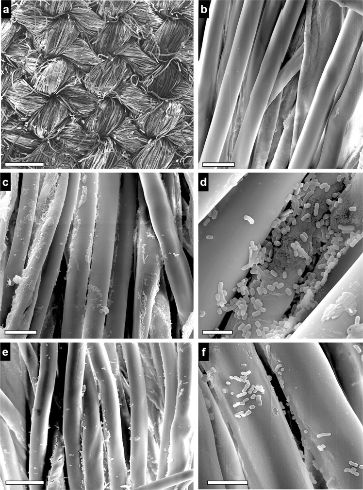
Fig 4. SEM analysis.

Scanning electron microscopy analysis of the morphology of A. baumannii (strain ATCC® 19606T) cells maintained on white lab coat fragments for up to 60 days. Panel shows random microscopy fields observed at different magnifications. a,b, control samples, and c,d, infected samples at the beginning of the experiments (day 0). e,f, infected samples after 60 days at 22ºC. Original Magnification: a, ×100; b,c,e, ×2.500; d, ×10.000; f, ×5.000. Scale bars: a, 0,5 mm; b, 20 μm; c,e, 25 μm; d, 5 μm; f, 10 μm.
