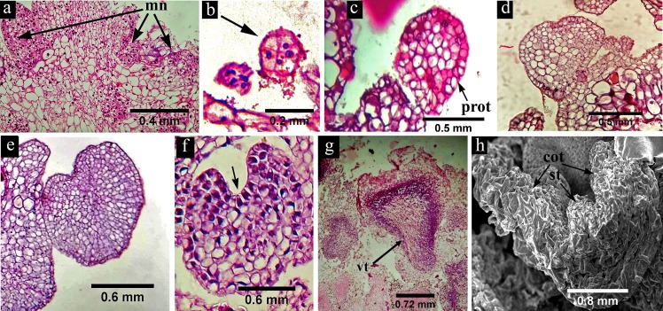
Fig 3. Histology and scanning electron microscopic view of somatic embryo development in H. sabdariffa var. HS 4288.
(a) The cross section of the embryogenic callus showing some meristematic nodules (mn) with darkly stained cells and prominent nuclei; (b) 8-celled pro-embryo (arrow mark) representing the first stage of somatic embryo initiation; (c) globular embryo with clear protoderm (prot); (d) early heart shape, (e) heart shape and (f) late heart shape somatic embryo with incipient cotyledon and clear cotyledonary notch (arrow mark); (g) torpedo shaped embryo with well demarcated vascular trace (vt); (h) germinating embryo with two cotyledons (cot) and elongated shoot tip (st).

