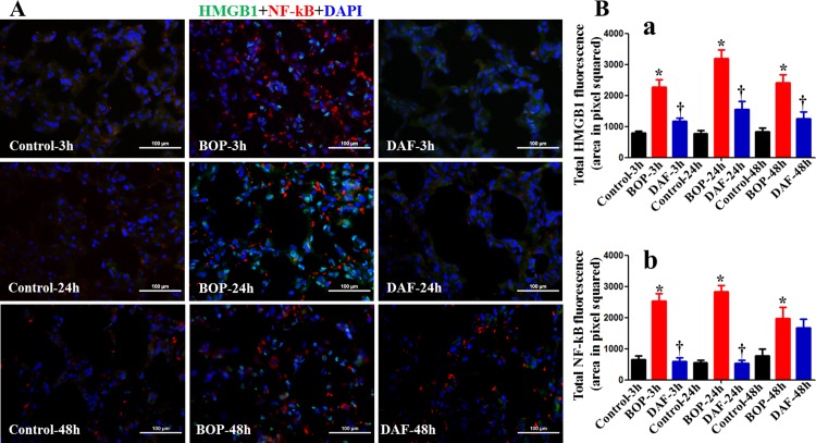
Fig 6. Effects of DAF on expression of HMGB1 and NF-κB in rat lungs exposed to BOP.
A) The expressions of HMGB1 (green) and NF-κB (red) in the lung tissue were measured by Immunohistochemical staining for the Control, BOP, and BOP with DAF treatment (designated as DAF) at 3h and 24 h after BOP, and representative images were presented, Original magnification = 400×, and scale bar = 100μm. B) Mean fluorescence intensities for HMGB1 (panel a) and NF-κB (panel b) from all the groups of control, BOP, and BOP with DAF treatment at 3h and 24 h after BOP were quantitated with criteria as described in Materials and Methods and graphed. n = 8 for all the groups except 48 h experimental samples (n = 5). Bar graph values were expressed as mean ± SEM, and compared using one-way ANOVA followed by Bonferroni test. C) Active caspase-3 was stained with anti-active caspase-3 antibody by IHC, and mean fluorescence intensities were calculated using the criteria as described in Materials and Methods, n = 5 for each group, and the data were expressed as mean ± SEM, and compared using one-way ANOVA followed by Bonferroni test.

