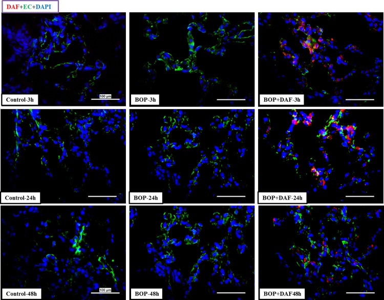
Fig 8. Deposition of rhDAF in rat lungs.
Representative micro-photos were from frozen sections of rat lungs immunostained with anti-human DAF and anti-endothelial cell antibodies. Original magnification = 400×, and scale bar = 100μm. n = 8 for all the groups except 48 h experimental samples (n = 5).

