
Figure 3. Clinical and genetic heterogeneity among nine subtypes of PFA ependymoma.
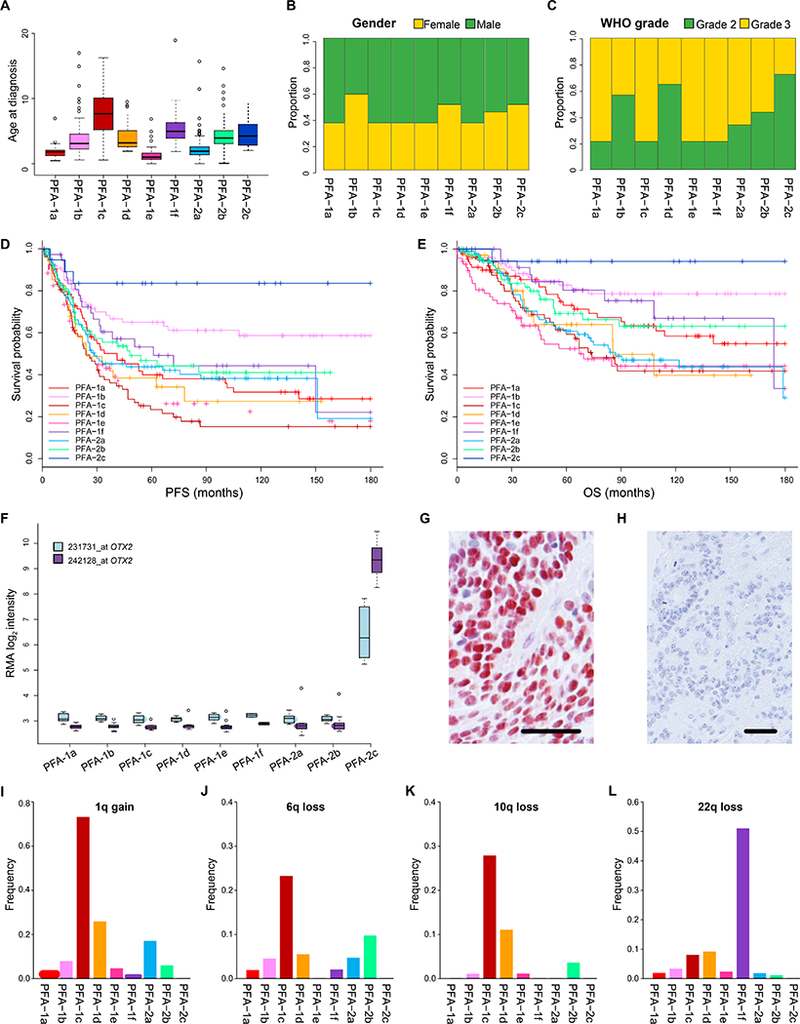
Nine subtypes of PFA ependymoma displaying variability in: (A) age at diagnosis (years), (B) gender ratio, (C) ratio of pathological grade, (D) progression-free survival (PFS), and (E) overall survival (OS). (F) Elevated OTX2 expression in PFA-2c ependymomas demonstrated by gene expression profiling (2x Affymetrix u133 v2 array probes) and immunohistochemistry (G). Immunonegative PFA ependymoma of another subtype (H). Varying frequencies across subtypes of the four commonest chromosome arm copy number alterations found in PFA ependymomas; 1q gain (I), 6q loss (J), 10q loss (K), 22q loss (L). Scale bars (G/H) = 100μm.