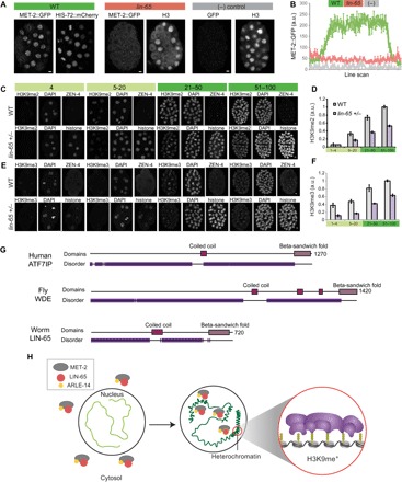
Fig. 6. LIN-65 is rate-limiting for H3K9me2 and nuclear MET-2 during embryogenesis.

(A) Distribution of MET-2::GFP in WT versus lin-65 mutants versus no-GFP WT strain. Scale bars, 2 μm. Note that this H3 antibody detects mostly cytosolic histone H3 during mitosis. (B) Line-scan analysis across embryonic nuclei shows mean MET-2::GFP intensity in WT (green) versus lin-65 (pink) mutants versus no-GFP control (“−,” gray). Average of line scans across multiple nuclei is shown, and error bars denote SEM. (C and E) H3K9me2 and H3K9me3 levels in embryos with a single-copy ZEN-4::GFP (“WT,” on-slide control) or progeny of lin-65(+/−) heterozygous mothers identified by HIS-72::mCherry at designated stages of embryogenesis. (D and F) Quantitation of H3K9me2 and H3K9me3 levels from WT (gray) or lin-65(+/−) (purple) offspring. Error bars denote SEM. (G) Domain architecture from HMMER (www.ebi.ac.uk/Tools/hmmer/) for human ATF7IP, fly Windei (WDE), and worm LIN-65 showing disordered (purple), coiled-coil (pink), and β-sandwich fold (violet) regions. (H) In early embryos, MET-2 (gray), LIN-65 (red), and ARLE-14 (yellow) are enriched in the cytosol, and there is little H3K9me2 or heterochromatin (light green). As embryos mature, MET-2 and interactors gradually accumulate in nuclei, form concentrated nuclear hubs, and deposit H3K9me2. MET-2–dependent H3K9me is required to generate heterochromatin domains (compacted, dark green).
