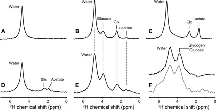
Fig. 1. In vivo 2H NMR.

Deuterium NMR spectra acquired without localization from (A) rat brain in vivo at 11.7 T before infusion of any 2H-labeled substrate, (B) rat brain after infusion of [6,6′-2H2]glucose in vivo, (C) rat brain after infusion of [6,6′-2H2]glucose postmortem, and (D) rat brain after infusion of [2H3]acetate in vivo. (E) 2H NMR spectrum acquired from human brain at 4 T, 60 min after oral administration of [6,6′-2H2]glucose. (F) 2H NMR spectra from rat (top, black) and human (bottom, gray) liver after intravenous and oral administration of 2H-labeled glucose, respectively. Each spectrum shown represents 1 min of signal averaging. While the lower magnetic field strength used for human studies leads to a proportionally lower spectral resolution [compare (B) and (E)], the sparsity of the 2H NMR spectra in combination with the absence of large water or lipid signals still allows robust signal identification and quantification (see also Figs. 2 to 5).
