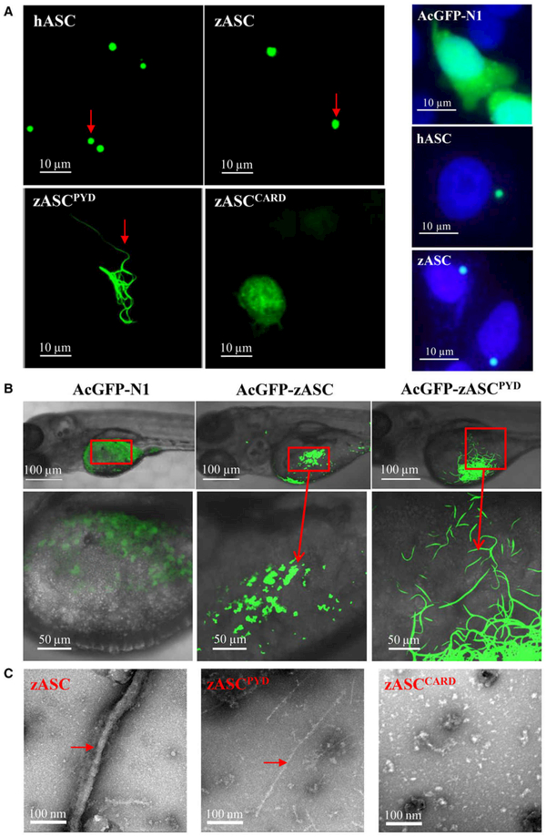
Fig. 2.
Both the PYD and CARD of ASC are required for speck formation. (A) Left side: subcellular localization of the AcGFP-fused hASC, zASC, zASCPYD, zASCCARD and AcGFP-N1 proteins. Bars: 10 μm. Right side: ASC SPECK formation close to nucleus per cell. 4’,6-Diamidino-2-phenylindole (DAPI) staining was used to label nucleus. (B) Representative confocal images of the zebrafish embryo yolk circulation region when zASC and zASCPYD are overexpressed. Bars: 50 and 100 μm. (C) Representative electron micrograph of the zASC, zASCPYD and zASCCARD proteins subjected to negative staining. Bar, 100 nm.

