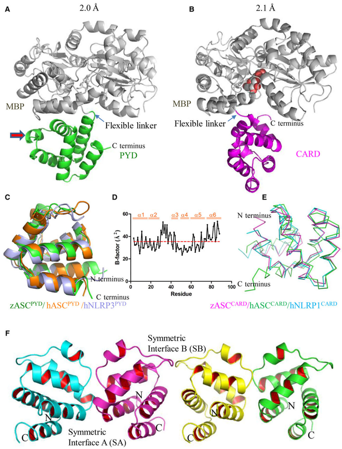
Fig. 3.
Crystal structure of zebrafish ASCPYD and ASCCARD at atomic resolution. (A) Crystal structure of the PYD of zASC (green) with an MBP tag (gray) at 2.0 Å resolution. The MBP protein was used as a crystallization tag to facilitate the formation of protein crystals. A maltose ligand was built in the ligand-binding pocket of MBP (same for B). (B) Crystal structure of the CARD of zASC (magenta) with an MBP tag (gray) at 2.1 Å resolution. (C) Structural superposition of PYD structures. The crystal structure of zebrafish ASCPYD (green) was superimposed on the NMR structure of human ASCPYD (1UCP, orange) and the crystal structure of human NLRP3PYD (2NAQ, light blue); the overlay yielded RMSD of 1.18 and 1.08 Å, respectively. (D) B-factor analysis of the zASCPYD structure. (E) Structural superposition of the CARD of zebrafish ASC, human ASC (2KN6, green) and human NLRP1 (4IFP, blue). (F) PYD–PYD interaction interface found in the crystal lattice.

