
Figure 5.
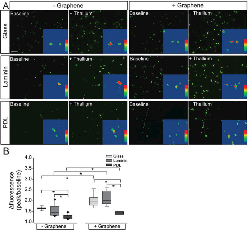
Cation channel activity in RGCs on culture substrates with graphene overlay. A) Representative fluorescent micrographs of RGCs cultured on glass (top), laminin (middle), or PDL (bottom) platforms with (+) or without (−) graphene overlay. RGCs were loaded with the cell-permeable dye Thallos (green). Images were taken at baseline and after addition of thallium, which binds to and increases the fluorescent intensity of Thallos dye. Scale bar = 100 μm. Insert: zoom of an individual cell within the larger image analyzed with a heat map showing the fluorescent signal of Thallos dye. B) Box plot of the change in the fluorescent intensity of each cell (peak intensity/baseline intensity).