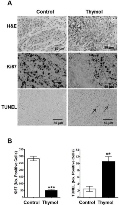
Fig. 3. Histopathological analyses of Cal27-derived tumors treated thymol.

A) Representative photomicrographs of Cal-27 derived tumors (40 × magnification) stained with H&E or Ki67, and TUNEL assays are shown; apoptotic cells are illustrated by arrows; scale bars = 50 μm. B) Quantification of Ki67 positive cells and apoptotic cells in treated tumors (mean ± SEM; n=3 per group; 3 fields per specimen; **p<0.01, ***p<0.001).
