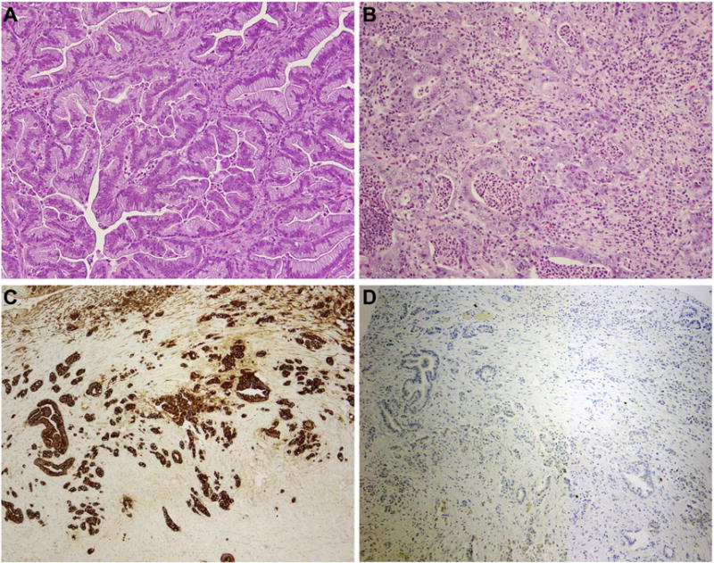
Figure 1. Histologic features of primary mucinous ovarian carcinomas.

Representative hematoxylin and eosin stained sections of (A) a mucinous adenocarcinoma with an expansile pattern of growth and (B) a poorly differentiated mucinous adenocarcinoma with destructive stromal invasion. (C) Poorly differentiated mucinous adenocarcinoma showing strong CK7 expression. (D) Poorly differentiated mucinous adenocarcinoma displaying lack of CK20 expression. Magnification 400x.
